| |
|
|
|
|
|
|
|
|
|
|
|
|
|
|
|
| • |
| • |
| • |
| • |
| • |
| • |
| • |
 |
|
|
|
| |
|
|
|
|
|
|
|
| HISTOLAB® : Get
access to EASY Image Analysis ! |
|
HISTOLAB® is a software designed to
give an easy access to image analysis tools for each of
you at the lab for routine work. Forget complex endless
menus, hard-to-read user's manuals and specialist's words
! HISTOLAB® allows you to do image analysis without
the help of a specialist. Capture, archive, improve, detect,
count, quantify, process, report and export....HISTOLAB®
is a package of the mostly currently used image analysis
tools at the Life Sciences Labs.
However if, despite our efforts, HISTOLAB® can't
fullfil your needs, don't hesitate to contact us, we will
be happy to develop for you dedicated
applications for increased performances and low investment
cost. |
 |
| (Click to enlarge) |
|
|
 |
| (Click to enlarge) |
|
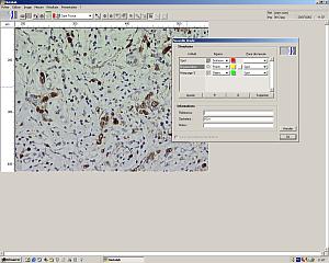
Define the measurements
With HISTOLAB®, you can quickly and simply define
what you want to analyse : zone of interest, cells, nuclei,
structures, make counting, measurements, quantitations...Each
measure will be associated automatically to the corresponding
object defined earlier for a facilitated results processing. |
|
 |
| (Click to enlarge) |
|
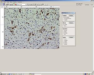
Define automated detection
protocol
A tutorial window will guide you, with simple mouse-clicks
and without programming, through the definition of the
detection protocol of the objects to be analysed. This
can be done either on an archived image or on a real time
video image, allowing you to select various areas of your
slide to improve the detection method. The detection protocol
can be saved for repeated use. When an automatic detection
can't work properly (too low contrast...), user may apply
manual measurement or counting. The results will be automatically
saved to a table in the same way as for the automatic
detection. |
|
 |
| (Click to enlarge) |
|
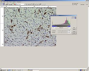
Improve images
HISTOLAB® provides a full palette of image improvement
functions. These tools may be used to improve the image
display for reporting reasons or to facilitate the automatic
detection of objects by, for example, improving the contrast
between objects to be measured and background. |
|
 |
| (Click to enlarge) |
|
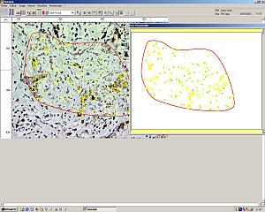
The automated detection
The automatic detection protocol may be applied to the
whole image or to a selected zone. Multiple zones (ex.
: normal tissu, pathologic tissu) may be selected and
have their own measurements realised. The detection protocol
can be applied to an archived image or a real time video
image thus allowing you to make multiple measurements
in various areas of your slide sample. Equally, working
on a real time video image saves space on disk as only
the data are recorded and not the images unless you want
to keep them.
If you upgrade your microscope with an encoded or motorized
stage, the software will recognize the actual position
on the slide thus eliminating the risk for duplicate measurements
on the same points. It also allows you to have a global
overview (see windows in the image above) of the measurements
made on your sample. |
|
 |
| (Click to enlarge) |
|
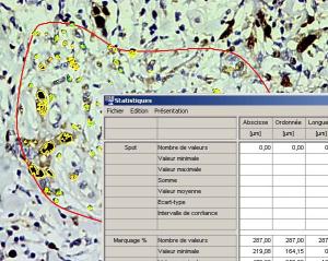
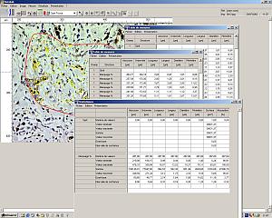
Processing the data
The results are stored automatically into tables, are
processed for statistics (average, standard deviation...)
and graphs generation of different types.
Each table and graph can be readily exported to an MS
Excel sheet for further processing or reporting. Captured
images can be associated to the tables and graphs.
For many samples, a scanning detection protocol can be
applied to automatically scan the sample and collect the
data.
Contact us for specific
applications. |
|
|
To order
HISTOLAB®
Catalog #
|
| |
|
|
|