|
|
|
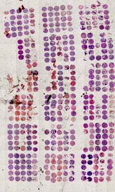
Tissue Array
|
|
|
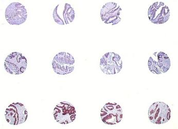
Tissue Arrays : hundreds
of tissue sections on one single microscope slide
The tissue array consists of building, starting from blocks
of tissues embedded in paraffin or frozen, new blocks
containing several tens to several hundreds of cores of
these tissues in order to be able, after cut of these
blocks of tissue arrays, to mount microscope slides displaying
several tens to several hundreds of tissues.
The tissue array constitutes a new powerful tool for studies
in post-genomic and post-proteomic validation.
You can build your own tissue arrays with the Tissue Arrayer.
Tissue arrays can be built starting from your own tissues
under service contract.
Some tissue arrays are also available on catalogue although
having a diversity and a richness less than the majority
of the tumour libraries. |
| |
|
Potentiate
your tumour library in the research programs
The transfer of your tumour library on tissue array allows
to increase considerably its potential of use and spreading.
Your tumour library is much more easily available in the
form of slides comprising whole or part of your tumour
library. |
|
|
You can quickly and simply
build various combinations of your tumour library
in order to answer various problems: pathology at
various stages, pathology at various individuals,
various ethnic populations, different populations
with therapeutic answer, various bodies at one or
more individuals, etc. The combinations are limited
by your experimental imagination and your needs.
You master the use of your tumour library perfectly.
Your blocks of tissues, rare and invaluable, remain
in your laboratory, you avoid the accidental losses.
The tissue array enables you to diffuse your tumour
library in the format of glass slides, easy to transport
and especially infinitely less consuming than using
the original blocks. You master the quality of the
tissues on which you will work. The tissue array
enables you to obtain great numbers of slides (see
Section Tissue Array section for more details) seriated
which will display very close characteristics. You
optimize your studies and facilitate the work of
your partners by focusing the analysis on the most
significant taking away in each one of your blocks
of tissues. The traceability of the slides is finally
easier to control than those of the blocks in the
majority of the cases, and particularly in the case
of old blocks files. |
|
|
Easily
consider studies on great numbers of tissues
The use of tissue arrays makes possible studies on very
large numbers of tissues without significantly expanding
or changing your lab organization. You can still continue
to use the same equipment and the same manpower.
Improve the statistical
significance of your research results
The use of tissue arrays in your research and validation
programs allows you to eliminate classical variation factors
like inter-slide, inter-staining bath or inter-operator
variability.
You can define experimental plans on very large numbers
of tissues to increase the statistical significance of
your results and avoid the mistake of conclusion based
on a particular case.
You can define experimental plans in which all tissues
to be compared are on the same slide. You can easily incorporate
controls for staining, pathology grades, etc.
The interpretation of your tissues is easier and more
rigourous. |
| |
Reduce
your response time
The tissue array allows you to simultaneously study multiple
cases whithin a very short time frame compared to classical
full section.
You can use tissue array for clinical studies or validations
of new markers for clinical routine like antibodies newly
introduced on the market. By this way, you can rapidly
obtain accurate responses and make decisions far earlier
to use new antibody for therapeutic treatment or routine
diagnostic.
Make considerable savings
on reagents
Protocoles that you classically use every day on your
slides can be applied readily on the tissue array slides
without modification. Do not change concentration nor
volume of marker poured on the slide. From then, savings
are considerable and directly proportionnal to the number
of tissues in your tissue array. Large cohorte studies
become possible with limited budgets. |
|
| Imagine all the possible applications
! |
| |
 |
Few examples :
- Validation of genes or proteins identified during genomic
or proteomic work ;
- Validation of new markers of tumour progression ;
- Development of new diagnostic IHC techniques ;
- Protein distribution studies in the whole set of body
organs ;
- Correlation studies of new markers during tumour progression
;
- Histological studies on large population ;
- Correlation studies between therapeutic response and
expression of some markers on a large population discriminated
in sub-populations on DNA chips ;
- Correlation between chromosomal aberrations and proteins
expression by IHC and FISH ;
Other ideas ? |
|
|
|
|
|